Virophage, Viroporins and Trogocytosis
Time for some microbiology,

Desnues C, Boyer M, & Raoult D (2012). Sputnik, a virophage infecting the viral domain of life. Advances in virus research, 82, 63-89 PMID: 22420851
Nieva JL, Madan V, & Carrasco L (2012). Viroporins: structure and biological functions. Nature reviews. Microbiology, 10 (8), 563-74 PMID: 22751485
Osborne DG, & Wetzel SA (2012). Trogocytosis Results in Sustained Intracellular Signaling in CD4+ T Cells. Journal of immunology (Baltimore, Md. : 1950), 189 (10), 4728-39 PMID: 23066151
Further reading
I'm simply amazed at the set of terminologies that keep arriving in microbiology. For last few days, I spent some time listening to some talks which i could grab hold of online. When i completed listening to the talks, i came to the conclusion that i could have a whole blog post just talking about some terminologies that have slowly managed to squeak into the Medical literature in terms of microbiology and immunology. One of them is vomocytosis, about which i have already written. More to come include- Viroporins, Virophage, Trogocytosis, NETosis, Paleovirology, Fungivore etc.
Virophage:
 |
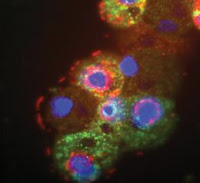
Photo 1: Giant mamavirus particles (red) and
satellite viruses of mamavirus
called Sputnik (green). Source
|
So what is this virophage? The name would imply to a reader almost immediately that these viruses that infect other viruses. But, the truth is this isn't true. Baffling!! Virophage is actually a misnomer. When the first giant Mamavirus was discovered (inside an Acanthameoba), a smaller co infecting virus referred as Sputnik was seen as a companion. Scientists soon realized that the sputnik was taking advantage of Mamavirus replication factory for its own replication. Moreover, presence of Sputnik reduced the turnover of Mamavirus!!
This observation went as far as to say that viruses can be infected by other viruses and hence virus should be considered to be a living entity. The comment by Jean-Michel Claverie, a virologist reads so “There’s no doubt this is a living organism. The fact that it can get sick makes it more alive”. The argument has taken a long way of impressive research. With current set of understanding, virophages looks more like, commonly known helper viruses. However, the exception is that in this case there is a actual viral interference. But i still have enough doubts to say one virus parasitize another, simply because the first and the second virus both use the same host resource, rather than the later using earlier virus entirely. This probably means the two virus competing with each other for the same niche.
Viroporins:
 |
| Fig 1: Viroporins, Source |
Viroporins are small proteins (typically 50- 120 amino acids long), produced by virus encoding a function of ion channel. The most important feature is that they contain at least one hydrophobic transmembrane, to form an amphipathic alpha helix. Based on the structure the viroporins are classified into 2 class- Class I (have a single membrane-spanning domain) and Class II (form helix–turn–helix hairpin motifs that span the membrane).
Some of the best studied viroporins include- Poliovirus 3A, Poliovirus 2B, Coxsackievirus B3 2B, HIV-1 Vpu, Influenza A virus M2, Influenza B virus NB, HCV p7, BEFV alpha-1 protein etc. A wide variety of functions are implicated to be caused by viroporins. This inculdes- Alteration of plasma membrane potential, Alteration of cellular Ca2+ homeostasis, induce intracellular membrane remodelling to generate viroplasm and essential part in assembly, budding and release of the viral progeny.
Trogocytosis:
 |
| Fig 2: Trogocytosis. Source |
When a cell, lends its membrane lipids and membrane bound proteins, directly from one cell to other, the phenomenon is called as Trogocytosis. This basically is a synapse between two cells (as shown in Fig 2 to the left) that leads to transfer of materials. The mechanism of extreme importance in immunology and occurs commonly than actually thought.
Trogocytosis has been shown to be important in transfer of antigenic material from macrophages to lymphocytes, uptake of macrophage Fc receptors and MHC molecules by T cells, acquisition of recipient
MHC class I and II molecules on donor thymocytes in bone-marrow chimaeras, transfer of MHC class II
proteins from splenic cells to allogenic T-cell clones, capture of B-cell surface immunoglobulin by T cells and more (Taken from Ahmed etal). More recently it has also been shown to be important in sustaining Intracellular Signaling in CD4 + T Cells
Desnues C, Boyer M, & Raoult D (2012). Sputnik, a virophage infecting the viral domain of life. Advances in virus research, 82, 63-89 PMID: 22420851
Nieva JL, Madan V, & Carrasco L (2012). Viroporins: structure and biological functions. Nature reviews. Microbiology, 10 (8), 563-74 PMID: 22751485
Osborne DG, & Wetzel SA (2012). Trogocytosis Results in Sustained Intracellular Signaling in CD4+ T Cells. Journal of immunology (Baltimore, Md. : 1950), 189 (10), 4728-39 PMID: 23066151
Further reading
1. Fischer MG, Suttle CA. A virophage at the origin of large DNA transposons. Science. 2011 Apr 8;332 (6026): 231-4. Link
2. Gonzalez ME, Carrasco L. Viroporins. FEBS Lett. 2003 Sep 18; 552 (1): 28-34. Link
3. Ahmed KA, Munegowda MA, Xie Y, Xiang J. Intercellular trogocytosis plays an important role in modulation of immune responses. Cell Mol Immunol. 2008 Aug;5(4):261-9. Link


Comments
Post a Comment