Lab series# 9- Pulsed field gel electrophoresis
Greetings,
Electrophoresis is a term that everyone has heard about. For people doing science in a wet lab setup, electrophoresis is kind of a usual work set up. There is a large number of variations of this technique, depending on the objective to be achieved. Gel electrophoresis is the one classic example. I haven't personally worked with a PFGE. Since I had to give a seminar on the basics of this topic, I did a little bit of literature reading. Here is the summary.
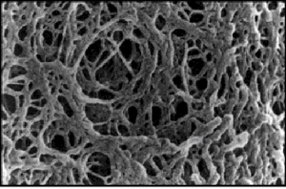 |
Photo 1: SEM image of
agarose polymer. Source
|
But there is a drawback to this "Conventional" technique. Larger the DNA fragment you have to analyse the more you need to loosen up the pore for DNA to move. This means, a more diluted gel needs to be made. There is a limit to how much diluted gel you can make and hence an inherent limit to the size of DNA that can be resolved. It has been calculated that the conventional gel electrophoresis can effectively separate DNA fragments up to ~20 kb, though practical resolvable limit is probably much less than this value. Any DNA fragment larger than this limit will stay at the same place regardless of DNA size, since the size is too large for it to pass through the gel pores. This is where the technique of PFGE comes in handy. PFGE methodology is useful when the analytical DNA sample is expected be of larger size (in Kbp lengths).
 |
| Fig 1: Schematic representation of PFGE work flow. Source |
So how does the PFGE basically work? PFGE was first demonstrated by Schwartz and Cantor (Link). The idea was based on a earlier observation that DNA molecules elongate upon application of an electric field and return to an un-elongated state upon removal of the electric field, the relaxation rate being dependent on the size of the DNA. Larger pieces of DNA will be slower to realign while smaller pieces will be quicker. Thus over a long run with consistent change of electric fields the larger DNA samples will migrate in the gel yielding a pattern which can be analysed.
Preparation of DNA sample for PFGE is slightly different from the conventional methodology. This is because handling large piece of DNA tends to be damaged easily. For bacterial typing the most recommended method is to use a fresh culture and embed the bacteria into a gel. This is known as the plug. Currently plug holds are commercially available. The cell lysis and restriction enzyme treatment is done on the plug. This plug is the loaded into the PFGE. A schematic representation of the PFGE- process is shown in Fig 1.
PFGE has multiple parameters that can be varied to achieve optimised results. Of all the variable parameters- Pulse time and reorientation angle appears to be the most crucial.
 |
| Table 1: Pulse time recommended for DNA fragments. Source |
Pulse time (also known as switch time or switch interval) is defined as time interval for which a given set of electric field is active. The larger the expected size of fractionated molecules, the less frequently the switching of electric fields should be performed. However, In a given pulse time only molecules of a particular size class occur under optimal conditions for separation and may be effectively fractionated. This means that the over a range of DNA size different pulse time needs to be used. The change of pulse time during electrophoresis is called “Ramping”. This parameter may be increased throughout a run either constantly or discontinuously over a range of discrete values. Continuous increase of pulse time from minimum to maximum of specified values during a given time interval is called “Interpolation”. An alternative variant, the stepwise increase in pulse time, is called “Stepping”.
The acute angle between the two alternating electric fields is called as reorientation angle. In certain instruments this is not variable and fixed at 120°. In some instruments this can be varied as per requirements. Reorientation angle does not produce significant effect in DNA molecules with less than 1 Mbp size. The mobility of larger molecules increases with decrease of the angle; But it has been the common experience by users that the band resolution is significantly reduced with decreasing angle. Lesser than 90° is not recommended. For a start 110° is used as optimal in most cases.
 |
| Table 2: Variants of PFGE. |
PFGE is a prototype version. The principle of this technique has been used in different instruments. Some of the most well known include- FIGE, TAFE, CHEF and RGE. See Table 2 for details.
CHEF is probably the most commonly used apparatus. The CHEF apparatus has twenty-four point electrodes equally spaced around the hexagonal contour and voltage is set at all points. It employs the principles of contour-clamped electrophoresis to generate homogeneous electrical fields by holding the electrodes at intermediate potentials, thus generating the homogeneous electric fields necessary for straight, distortion-free lanes. CHEF uses a fixed reorientation angle of 120° with gradiations of electropotential radiating from the positive to the negative pores. This allows resolution of DNA fragments up to 7 Mbp.
PFGE technique is currently considered as a gold standard for epidemiology of bacterial infection outbreaks. The resolution provided is quite high in comparison to other methods such as Ribo-typing or MLST. However, there is a lack of consensus on protocol for a variety of species and for some bacteria PFGE cannot be performed. Inter-laboratory variations have been another issue. Experts agree that PFGE gives a phylogenetic map but it should be considered as a relative map rather than absolute. According to me the only technique that is better is Whole genome sequencing.
PFGE technique is currently considered as a gold standard for epidemiology of bacterial infection outbreaks. The resolution provided is quite high in comparison to other methods such as Ribo-typing or MLST. However, there is a lack of consensus on protocol for a variety of species and for some bacteria PFGE cannot be performed. Inter-laboratory variations have been another issue. Experts agree that PFGE gives a phylogenetic map but it should be considered as a relative map rather than absolute. According to me the only technique that is better is Whole genome sequencing.

Sambrook J, & Russell DW (2006). Pulsed-field Gel Electrophoresis via Transverse Alternating Field Electrophoresis Gels. CSH protocols, 2006 (1) PMID: 22485453


Comments
Post a Comment