Lab series# 15: Biochemical tests for identification of bacterial isolates
Classic clinical microbiology techniques such as culture and phenotypic analysis form the major chunk of microbial identification, especially so for the bacterial isolates. The most sophisticated microbial laboratories still use the microbial culture techniques and need to isolate the bacteria and biochemically identify the isolate. Many different automated biochemical testing equipment are available which uses the same principle, only that the system is automated. In a majority of the microbial laboratories around the world, molecular tests are not available or feasible and identification is made through classic biochemical tests.
There are a large number of biochemical tests at the disposal of a microbiologist, but the choice of the panel of tests is based on preliminary findings such as gram staining pattern and growth characters which hint to what the list of organisms can be. For example, finding a gram negative bacilli growing in McConkey agar from stool sample would hint looking at Enterobacteriaceae group and tests like coagulase (which is used for identification of coagulase producing staphylococcus species) would of be no use. In most common scenario less than 15 biochemical tests are required for reliable identification of a bacteria to species level. Having more biochemical tests can increase the confidence in identification, but performing every possible biochemical test is counter productive.
Phenotypic-biochemical tests can be classified into 3 groups
1. Universal
These tests are done for almost any isolate and guide the microbiologist to a possible set of biochemical tests that needs to be done to get a reliable identification.
Examples: Hemolysis pattern, Motility test, Catalase test, Oxidase test
2. Differential
These are a common set of tests that are done to identify the isolate up to species level. The identification is made based on the results from a combination of tests and individual results by themselves are not sufficiently informative to make an identification.
Examples: IMViC tests, Triple Sugar Iron test (TSI), Sugar utilisation tests
3. Specific
These are tests that are specific to a particular set of species or for sub-typing a species. These tests are usually performed to confirm or identify at the subspecies level. The individual tests are informative by themselves in this case.
Example: γ-Glutamyl aminopeptidase test
It should be noted that certain tests are combinatorial in nature and there is more than one test to assess the same phenotypic parameter. For example, Mannitol Motility test is a combination test. The test can assess motility and the ability to utilise Mannitol. Motility can also be tested by using hanging drop technique and mannitol can be assessed using sugar utilisation tests.
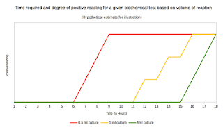
 In earlier days, most tests were done in large volumes typically using more than 5ml of a medium which takes more time to produce a visible result. Let us take an example case of lactose utilisation. If I’m to put an E coli into 5ml of a lactose-containing medium, it would take the isolate more time to break down lactose and give a pH change sufficient enough to give a reading in comparison to doing the same in a 0.1 ml medium. The simplest way to minimise the time involved in reading it to reduce the total reaction volume, which is one of the key concept in the most automated system allowing faster reading of results. Fig 1, is a hypothetical graph showing the time required to give a positive result in different conditions (The exact details will change depending on the test and other conditions).
So let us talk about few tests of interest in detail. The procedure for these tests can be found anywhere and hence the preparation of the medium and SOP for testing is not discussed. I want to focus on understanding the test principle.
1. Hemolysis Pattern on Blood agar:
 |
| Table 1: Hemolysis patterns that are seen in blood agar. |
It should be noted that hemolysis is dependent on the enzyme involved and variations are seen for the same species depending on conditions. A study by Vancanneyt et al which tested multiple strains of E faecium found the following. beta-hemolysis was observed on both sheep and human blood for only 1 strain, 4 strains showed beta-hemolysis on human blood, but not on sheep blood, and rest of the strains studied showed no hemolysis on either medium. Of course, beta hemolysis is not a common feature for Enterococcus group.
 |
| Photo 1: Blood agar Plate. Source |
It has been noted in certain labs that human blood is used for the preparation of blood agar. Standards recommend that this should not be practised since the human blood may contain bloodborne pathogens which form a risk to the technical staff preparing the agar and also the blood may contain inhibitors which may hamper growth. Some groups have questioned this thinking. In reality, human blood for the preparation of blood agar is usually obtained from blood bank stored bags which have reached expiry date or has expired. The blood bank usually tests such blood samples for common TTIs (Transfusion transmittable infections) and hence the risk of using such samples is minimal. Since the application is to grow human pathogens they should, in theory, be able to overcome inhibitors if any and hemolysis obtained is significant since it closely mimics human scenario. A set of counter argument is that using blood samples from humans that have expired is likely to have degraded and hence, false results would be common.
Overall, it is recommended that sheep blood is used for best results and the sheep which is used for obtaining blood be occasionally tested for potential infection. Currently, most laboratories around the world rely on commercial vendors to supply the lab with readymade disposable sheep blood agar plates that are quality controlled, thus avoiding the hassles.
2. Catalase test:
Catalase is an enzyme produced by a few group of bacteria and their primary function is to neutralise hydrogen peroxide activity abundantly expressed by attacking immune cells.
 |
| Fig 1: SOD and Catalase activity. From Prescott Textbook 5th Ed |
Catalase test is of 3 types-
- Qualitative catalase
- SQ (Semi Quantitative) catalase test
- 68 C (Heat stable) catalase test
In routine microbiology, qualitative catalase is the most commonly performed test and hence conventionally called as “catalase test”. The other two variants of the test are performed for selective mycobacterium isolates.
Catalase test is a test for demonstrating the presence of catalase enzyme by decomposition of hydrogen peroxide to oxygen and water.
 |
| Photo 2: Catalase test. Source |
Pseudo-catalase reactions are false positive reactions. They can be identified by their weak and late reaction and seen in some cases of Aerococcus species.
Semi-Quantitative catalase and 68C catalase test are specific tests used for differentiating mycobacterium species.
Most Mycobacterium species possess catalase which differs by quantity and heat lability at 68 C. SQ catalase reagent contains 10% tween 80 and 30% H2O2 LJ medium is inoculated with test organism and incubated for 2 weeks at 37 C and then tween-hydrogen peroxide reagent is added and allowed to stay for 10 min. The height of bubble formation is measured and reported as <45mm or >45mm. M kansasii, M simiae, and most scotochromogens give >45mm. M avium complex, M xenopi, M gastri etc give <45mm.
Certain Mycobacterium loses its catalase activity when suspended in a pH of 7 at 65 C for 20 min. M tuberculosis, M bovis, M hemophilum etc possess heat labile catalase. The test is most useful in differentiating member of Nonchromogenic mycobacterium.
3. Oxidase test:
 |
| Photo 3: Oxidase Test. Source |
Classically, 1% tetra-methyl-p-phenylenediamine dihydrochloride is freshly prepared daily and impregnated into a filter paper and dried. The colonies are smeared on the paper and look for colour change within 10 sec. Commercially available discs are now available which contains N, N-dimethyl-p-phenylenediamine oxalate, ascorbic acid and α-naphthol, a combination which is more stable thus avoiding the requirement of daily preparation.
Modified oxidase test is a special test used only for Gram positive, catalase positive cocci. It is commonly referred as Microdase test. Micrococcus oxidase enzyme is not readily accessible for reaction. This problem is overcome by the use of DMSO which permeabilizes the cell and permits access of reagents to oxidase enzyme.
Modified oxidase test is a special test used only for Gram positive, catalase positive cocci. It is commonly referred as Microdase test. Micrococcus oxidase enzyme is not readily accessible for reaction. This problem is overcome by the use of DMSO which permeabilizes the cell and permits access of reagents to oxidase enzyme.
4. Oxidative fermentation test:
The test was invented by Hugh and Leifson and thus sometimes also known as Hugh- Leifson Test or OF- glucose test. Bacteria can degrade glucose in a fermentative or oxidative manner. In either of the case, the end products are a mixture of acids which is indicated by an indicator. Bromothymol blue or Bromocresol purple are commonly used indicators. Bromothymol blue has a pH range of 6.0 - 7.6 and Bromocresol purple has a pH range of 5.2-6.8, both of which gives yellow colour in the acidic range. The medium contains a high concentration of carbohydrate and low concentration of peptic digest which reduces the possibility of utilising peptic digest to produce an alkaline condition which masks the acidity produced. The agar concentration is also kept low, which enables the determination of motility. Careful observation of the medium for breaks or rise in the medium can also be used to indicate gas production.
 |
| Photo 4: Oxidative-fermentative (OF) test. Source |
The test uses 2 tubes both containing OF medium and inoculated with bacteria. One is covered with a sterile mineral oil. This keeps the tube in an anaerobic condition. The tubes are then incubated for 24–48 hours. If the medium in the anaerobic tube turns yellow, then the bacteria are fermenting glucose. If the tube with oil doesn't turn yellow, but the open tube does turn yellow, then the bacterium is oxidising glucose. If the tube with mineral oil doesn't change, and the open tube turns blue, then the organism neither ferments nor oxidises glucose. Instead, it is oxidising peptones which liberate ammonia, turning the indicator blue. Motility can be observed in the medium by looing for growth trail. It should be noted that there are certain bacteria that take their own time to work the process and hence the test is not ideally read negative before 5 days of incubation.
Modified OF tests are used in special circumstances. For example, for testing halophiles, the OF medium is integrated with high salt concentration. There are certain bacteria that prefer other sugars, instead of glucose in which case OF medium containing other sugars can be prepared. For testing staphylococcus and micrococcus, Baird-Parker modification of the medium is recommended.
5. IMViC test:
 |
| Photo 5: IMViC test for E Coli. Source |
- Indole test
- Methyl Red test
- Voges Proskauer test
- Citrate utilisation test
Indole is an aromatic heterocyclic organic compound with a six-membered benzene ring fused to a five-membered nitrogen-containing pyrrole ring, which is derived from tryptophan using the enzyme tryptophanase. Most people recommend peptone water for testing indole production. Peptone water basically consists of Peptic digest of animal tissue and sodium chloride. A peptic digest is obtained by acid hydrolysis or enzymatic digestion. Acid hydrolysis method is harder and tryptophan is usually lost or reduced to very low levels by this method. The enzymatic method is much milder and uses trypsin and chymotrypsin combination. In each case, there is some loss of tryptophan and reduced availability of free tryptophan. Since most of the commercially available peptone is an enzymatic digest, peptone water should still work. However, if the nature of peptone is not known or results are not good it is recommended to add 1% tryptophan to peptone water and used for detecting indole.
Indole is a gas and can be detected easily with many different reagents. The most commonly used include Ehrlich's or Kovacs. Kovac’s reagent consists of para-dimethyl amino benzaldehyde in isoamyl alcohol and concentrated HCl. Ehrlich’s reagent uses Ethanol instead of Isoamyl alcohol and is more sensitive in detecting indole production especially in anaerobes and non-fermenters. Another sensitive alternative is p-Dimethylaminocinnamaldehyde (DMACA) in acidic solution impregnated into a filter paper, used as a spot test.
Methyl Red (MR) test:
 |
| Fig 2: The Embden-Meyerhof pathway for glucose dissimilation. |
Bacteria can utilise glucose through Embden-Meyerhof fermentation pathway and get into one of the end products, depending on the species. It can either produce homolactic acid or mixed acids containing a combination of lactic acid, acetic acid, formic acid, succinate and ethanol and gas formation if the bacterium possesses the enzyme formate dehydrogenase, which cleaves formate to the gases. Certain species can further get down the pathway and form 2, 3 butanediol from the condensation of 2- pyruvate. See Fig 2 for details.
The test is done on glucose phosphate peptone water. The MR test looks for the ability of bacteria to produce large amounts of acid resulting in significant decrease in the pH of the medium below 4.4. This acidic nature is indicated by methyl red (p-dimethylaminoaeobenzene-O-carboxylic acid) indicator which is yellow above pH 5.1 and red at pH 4.4. The test should be ideally read at 48 hrs since the test looks for sustained pH.
Voges-Proskauer (VP) test:
VP test is actually an extension of MR test and looks for the ability to produce butylene products. Acetoin (3-hydroxybutanone) is an intermediate in the reaction which is looked for using 40% KOH and alpha-naphthol. If acetoin is present, it is oxidised in the presence of air and KOH to diacetyl which reacts with guanidine components of peptone, in the presence of alpha- naphthol to produce a red colour. The test is read along with MR test.
Citrate test:
 |
| Fig 3: Citrate utilisation pathway. Source |
The test looks for the ability of a bacteria to utilise citrate as a sole source of carbon. For the bacteria to be able to do so, it requires 2 components- Citrate permease and citrate lyase. Citrate permease is a group of uptake proteins that allows the cell to uptake citrate and then lyase which converts citrate to oxaloacetate and acetate. The oxaloacetate is then metabolised to pyruvate and CO2.
The organism is inoculated into Simmon's or Koser's citrate medium. Simmons citrate agar contains sodium citrate as the sole source of carbon, ammonium dihydrogen phosphate as the sole source of nitrogen, other nutrients, and the pH indicator bromothymol blue. The bacteria converts the ammonium dihydrogen phosphate to ammonia and ammonium hydroxide, which creates an alkaline environment in the medium. At pH 7.5 or above, bromthymol blue turns royal blue which is otherwise green. Most people make the mistake of reading the reaction by colour. In some cases, the alkalinization doesn't occur (Or takes longer time) but colonies can be seen. Colony formation should be taken as an evidence of growth which is a reflection of the ability of the bacteria to utilise sole carbon source.
6. Triple Sugar Iron Test:
Triple sugar Iron is a complex test with multiple readouts. The test was first designed proposed by Sulkin and Willett which was later modified by Hajna for identification of Enterobacteriaceae members. The test medium is TSI (Triple sugar Iron agar). The test medium contains 3 sugars- Glucose (0.1%), lactose and sucrose (1% each). Phenol red serves as the indicator. The medium contains a butt and a slant. Ferrous sulphate serves as an indicator for H2S production. The medium is inoculated with a stab method on the butt and stroke method on the slant. The lower portion of the butt acts as an anaerobic condition since it is nearly inaccessible.
The first thing that happens when bacteria is inoculated, is to utilise the glucose. The amount of glucose is purposefully kept low (Nearly 10 times less in comparison to other sugars). If the organism can metabolise glucose in anaerobic conditions and aerobic conditions both the butt and slant becomes acidic turning the colour of indicator to yellow. This happens within 6-8 hours of inoculation. If the bacteria can utilise lactose or sucrose (or both), the acidification of medium continues and the medium remains yellow. If it cannot, the bacteria starts utilising amino acids by decarboxylation of peptone making the medium alkaline thus reversing the first acidic step. This gives a more reddish appearance. Phenol red has a pH range from 6.8 (yellow) - 8.2 (red). If the bacteria is a strict aerobe (ex Pseudomonas aeruginosa) the reactions occur only in the slant and the butt remains no change or non-reactive. If the bacteria is a facultative anaerobe, the reaction will be seen in both butt and slant. In general, more amounts of acids are liberated in butt region (fermentation) than in the slant (respiration).
Production of gas is evidenced by breaks or rising of the agar medium. Thiosulphate is reduced to H2S by several species of bacteria which combines with ferric ions of ferric salts to produce the insoluble black precipitate of ferrous sulphide. Reduction of thiosulphate proceeds only in an acid environment. There are several combinations of reactions possible that can be read. Following are the most common.
 |
| Fig 4: TSI reaction readouts. Source |
- The organism ferments glucose but does not ferment lactose or sucrose. The slant becomes red and butt remains yellow. It is reported as K/A (Alkaline slant/Acid butt)- remember butt is more acidic.
- The organism in addition to glucose ferments lactose and (or) sucrose. The slant and butt remain yellow. It is reported as A/A (Acid slant/Acid butt).
- If the organism is non-fermenter, Instead of sugars, peptone is utilised as an alternate source of energy under the aerobic condition on the slant which makes it alkaline indicated by the red colour while there is no change in the colour of the butt. It is reported as K/NC (Alkaline slant/No change)
In addition to the above gas and H2S is reported. Reactions in TSI should not be read after 24 hours of incubation because eventually sugars will be exhausted and decarboxylation reactions will take over making the medium alkaline.
The test cannot differentiate between lactose and sucrose fermenters. For this, a modification called as Kligler Iron Agar (KIA) which combines features of Kligler's Lead Acetate medium and Russell's Double Sugar Agar can be used. This medium doesn't have sucrose.
 |
| Fig 5: Fermentation test. Source |
7. Carbohydrate fermentation test:
The test is usually done on a Carbohydrate Fermentation Broth (Contains trypticase, Sodium chloride, and Phenol red) with 1% sugar which is to be tested. A durham's tube is kept in an inverted position which accumulates gas in case of gas production. The phenol red indicator turns yellowish if there is fermentation leading to acidic pH change. Alternatively, Andrade's indicator may be used.
Most often, a single sugar may not be sufficient enough to make a distinction and combination of multiple sugars are used. The most commonly used include- lactose, sucrose, xylose, mannose, arabinose, trehalose and maltose etc.
8. Urease test
Urease is an enzyme belonging to belong to the superfamily of amidohydrolases and phosphotriesterases. It catalyses the hydrolysis of urea into ammonia and Carbon dioxide.
- (NH2)2CO + H2O → CO2 + 2NH3 The formation of ammonia causes alkalinization of the medium, and the pH change is indicated by a change to pink at pH 8.1. Certain organisms can rapidly hydrolyze urea and their speed of hydrolysis can indicate the organism. A test called the CLO test (Campylobacter-like organism test), is a rapid urease test for diagnosis of Helicobacter pylori). A biopsy of mucosa is taken from the antrum of the stomach and is placed into Urea broth. A positive test in less than 30 min may be obtained indicating the pylori infection. Another method called Urea breath test is based on a similar idea but detection is based on isotope measurement.
- As already mentioned there are so many Phenotypic biochemical tests that can be performed for identifying an organism. However, with some experience and training, most organisms can be identified with few tests mentioned above at least up to the genus level.


Comments
Post a Comment