Understanding Autophagy: Nobel winning concept
Greetings
The recent announcement of Nobel prize 2016 in Physiology or Medicine to Yoshinori Ohsumi is perhaps a topic trending online these days. Autophagy is a physiological process, innate to a cell and has multiple implications in immunology and infectious diseases biology. I decided to step in and write some details of interest. Autophagy (or sometimes also referred to as autophagocytosis) is defined as a physiological tightly regulated, destructive mechanism of the cell that disassembles unnecessary or dysfunctional components. It is important to note that unlike other cellular degradation machinery, autophagy removes large macromolecular complexes and organelles that have become obsolete or damaged. It also has a primary role in clearing invading microorganisms and toxic protein aggregates.
 |
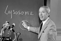
| Photo 1: Dr Christian de Duve. Source |
The roots of understanding autophagy come from an interesting observation by Christian de Duve studying the distribution of acid phosphatase but failed to detect any enzymatic activity in freshly isolated liver fractions. But the enzymatic activity reappeared upon storage for 5 days in a refrigerator. It was later understood that the proteolytic enzymes were sequestered in a membrane structure called the lysosome. Subsequent studies showed that in some cases they could also co-localise other cellular organelles with the lysosome. It became evident that the structures had the capacity to digest parts of the intracellular content and coined the term autophagy in 1963. There was some evidence that the process might have a role in human diseases, but nothing about the mechanism was understood.
 |
| Photo 2: Yoshinori Ohsumi. Source |
Autophagy is a complex pathway mediated by multiple different proteins. Though the pathway operates on a general principle, they are divided into 3 major pathways based on the specifics of the mechanism
1. Macroautophagy
2. Microautophagy
3. Chaperone mediated autophagy
Macroautophagy is the process by which the substrates are sequestered within cytosolic double membrane vesicles termed autophagosomes. In Microautophagy, a cytoplasmic material is trapped in the lysosome/vacuole by membrane invagination. It is especially important for the survival of cells under starvation.
Macroautophagy is the process by which the substrates are sequestered within cytosolic double membrane vesicles termed autophagosomes. In Microautophagy, a cytoplasmic material is trapped in the lysosome/vacuole by membrane invagination. It is especially important for the survival of cells under starvation.
 |
| Fig 1: Types of Autophagy. Source |
Chaperone-mediated autophagy (CMA) refers to targeting of proteins from the cytosol to the lysosomal membrane and then gaining access to the lumen by directly crossing its membrane. Proteins that undergo degradation by this pathway are identified through a recognition motif (such as amino acid sequence KFERQ motif). This allows for the removal of specific proteins thus becoming an efficient system for degradation of damaged or abnormal proteins. CMA is a multi-step process that involves following steps
- Substrate recognition and lysosomal targeting
- Substrate binding and unfolding
- Substrate translocation
- Substrate degradation in the lysosomal lumen
In general, Autophagy can be divided into multiple steps. Though most of these details have been worked on yeast models, mammalian genes have analogue proteins serving similar functions. Considering that the process is evolutionarily conserved the details are not expected to deviate much. The whole process in its simplest form can be described as forming a vesicle containing contents to be degraded and fusing a lysosome into it. The enzymes then degrade everything inside the pouch and contents are available for recycling. It is not clear, what are the exact signals that activate the process, but stress is known general factor.
 |
| Fig 2: Generalised autophagic pathway. Source |
Fig 2, is a simplification of Major events in Autophagy pathway. The process of autophagy involves the following steps
Autophagosome nucleation
 |
| Fig 3: Proposed process of Vesicle formation. Source |
This step is also known as Initiation. The initiation of autophagy can be triggered by a variety of extracellular signals which includes nutrient starvation, stress, microbial infection, toxins etc. An important target of these signals is TOR (Target of Rapamycin), a kinase that inhibits the autophagic pathway until this protein is inactivated by dephosphorylation. Class III PI3Ks are also good inducers of autophagy activation. The first step is to form a phagophore or isolation membrane. There are multiple proteins that are involved in this process, and they are called as ATGs (Autophagy-related proteins). Atg5–Atg12–Atg16 complex is recruited to the sequestration crescent, a double-membrane-bound structure that engulfs cytosolic constituents to become the closed, double-membrane-bound autophagosome. The process of vesicle formation is shown in Fig 3.
Growth and completion
The first step is to add phosphatidylethanolamine to Atg8. The carboxy-terminal amino acids of Atg8 are cleaved by cysteine protease Atg4 thereby leaving a conserved glycine residue. Cleaved Atg8 is then transiently linked to the Atg7 protein, then to Atg3, and finally to phosphatidylethanolamine. Modified Atg8 remains associated with autophagosomes until destruction at the autolysosomal stage. Cleaved Atg-8 enables fusion of autophagosome with a lysosome.
Autophagosome target and fusion
Autophagosomes fuse with endosomal vesicles mediated by small GTPases, such as the RAB proteins and acquire LAMP1 and LAMP2. These structures fuse with lysosomes and acquire cathepsins and acid phosphatases to become mature autolysosomes.
 |
| Table 1: ATG functional groups. |
Since one of the functions of autophagy pathway is to clear intracellular pathogens, inhibition of the pathway becomes important for the survival of the pathogen. Many different microbial proteins can achieve this goal. For example, Herpesvirus US11, Vaccinia E3L, Influenza virus NS1 inhibit PKR activation by dsRNA; HIV TAT forms complex with PKR, inhibiting its kinase activity; Legionella pneumonia delays acquisition of LAMP1; M tuberculosis blocks phagosome maturation. Intracellular microbial pathogenesis is full of such examples. Degradation of microbial proteins by autophagy are the source of peptides for presentation to the immune system.
Jeremy Berg captures the importance of Nobel award to workings of Ohsumi, "The process of autophagy that he discovered is now part of the fabric of modern cell biology and medicine. Researchers now consider defects in this pathway when trying to understand diseases." Ohsumi comments, "The human body is always repeating the auto-decomposition process or cannibalism, and there is a fine balance between formation and decomposition. That's what life is about.” He also noted that even though he has been studying the mechanism of autophagy for more than 27 years, he still doesn't think he fully understands it and hopes he could learn more.
References
1. Glick D, Barth S, Macleod K. Autophagy: cellular and molecular mechanisms. The Journal of Pathology. 2010;221(1):3-12.
2. Kaur JDebnath J. Autophagy at the crossroads of catabolism and anabolism. Nature Reviews Molecular Cell Biology. 2015;16(8):461-472.
3. Kirkegaard K, Taylor M, Jackson W. Cellular autophagy: surrender, avoidance and subversion by microorganisms. Nature Reviews Microbiology. 2004;2(4):301-314.


.svg.png)
Comments
Post a Comment