Lab Series# 4: Antigen Antibody reaction
Greetings,
Antigen- Antibody reaction forms the basis for most the laboratory workup methods, in the current scenario. Reactions come by several methodologies and are named accordingly. However, they all are built on the same basic rules and principles of reaction. In this post, we will explore a little bit of the basics ad talk about ELISA as a best classic example of Ag-Ab reaction.
Antigen- Antibody reaction forms the basis for most the laboratory workup methods, in the current scenario. Reactions come by several methodologies and are named accordingly. However, they all are built on the same basic rules and principles of reaction. In this post, we will explore a little bit of the basics ad talk about ELISA as a best classic example of Ag-Ab reaction.
There are 7 rules that defines all Antigen-Antibody reactions. Take any Antigen-Antibody reaction and the principle would belong to combination of rules laid below.
1. The reaction is specific, an antigen combining only with its homologous antibody and vice versa. The specificity, however, is not absolute and cross reactions may occur due to antigenic similarity or relatedness. The phenomenon was known as cross re-activity.
2. Entire molecules react and not fragment. When an antigenic determinants present in a large molecule or on a carrier particle reacts with its antibody, whole molecules or particles are agglutinated.
3. There is no denaturation of the antigen or the antibody during the reaction.
4. The combination occurs at the surface. Therefore, it is the surface antigens that are immunologically relevant. Antibodies to the surface antigens of infectious agents are generally protective.
5. The combination is firm but reversible. The firmness of the union is influenced by the affinity and avidity of the reaction.
6. Both antigen and antibodies participate in the formation of agglutinates or precipitates.
7. Antigens and antibodies can combine in varying proportions, unlike chemicals with fixed valencies.
Antigen + Antibody--------->Antigen-antibody complex
 |
| Fig 1: Ag-Ab reaction equilibrium |
 |
Table 1: Factors effecting equilibrium constant
|
 |
Fig 2: CDR in antibody that is involved
in binding to Antigen. Source
|
Immediately the question is raised what about the cross reaction? Consider 2 antibodies that fit onto an eptiope well. Suppose for argument sake there is an antigen and 2 antibodies (See Fig 3, shown below. Only the binding area is represented as linear diagram to simplify understanding).
 |
| Fig 3: Antigen and Antibody with charge configuration |
This raises a yet another important question. How much of such charge discrepancy a reaction bias can handle? The answer for this can be well understood with a mathematical model. Its a universal fact that the force of repulsion is always more than force of attraction (The analogy is "If you love someone, you love. But if you hate someone, you hate them from the core"). The equation reads so
B= A2- R4
In the equation B represents binding, A represents affinity and R is Repulsion. In Fig 3, there are a total of 7 points of interaction. Of theses 6 points of interaction represents "Affinity" and 1 point represents "Repulsion". So the equation dictates that B= 62- 14 which is a positive value. If the value is positive it means the binding would happen. If the equation yields Zero or negative value then the reaction wouldn't happen. That means, if there would be a discrepancy in 3 points in the above said reaction then the equation looks B= 42- 34 which is negative value. Oh yes, this is a total oversimplification as there are several points of interaction but serves the theoretical point of understanding. My point is, the total number of points interacting and how it interacts has the final say.
Last point, higher the negative value obtained (from above equation), higher is the repulsion. Higher the positive value obtained better is the binding. So if there is very small positive value the binding kinetics would still be poor.
There are several sets of antigen-antibody reaction such as agglutination, precipitation, ELISA, CLIA, Blot assays etc. Let us take ELISA as an example to talk about.
ELISA stands for Enzyme linked immunosorbent assay. The methodology was first described in 1971 by Engvall and Perlmann. The method seemed to so useful and convincing, almost any substance that can be antigenic and any antibody could be detected or even quantified with little tricks. The method ruled the field of wet lab science for almost 3 decades now and still continues to be so. Though many of the tests are now modified towards Chemiluminesence Immunoassay (CLIA), in most of the parts of the world, ELISA dominates.
 |
Photo 1: Dr. Eva Engvall
|
So what is this ELISA? The technique is a sub type of more broader group called as EIA (Enzyme immuno-assays). The ELISA technique uses a solid phase to immobilize the Ag-Ab complex and hence the name. Antigen and antibody specificity is used as the basis for the reaction. The reaction is signaled by the use of enzyme as marker, that can convert a substrate into colored compounds.
There are a wide array of solid phase that can be used for ELISA. The solid phase used can be classified into high capacity (Agarose, Sephadex, Cellulose, Nitrocellulose etc) and Low capacity types (Polystyrene, Polyvinyl chloride, Nylon, Glass). The capacity of solid phase can be manipulated as demanded by the assay. For instance, Cyanogen-Bromide activated Sephadex is a high capacity solid phase that has established binding with a wide range of proteins. It can be used in detection of IgE antibodies even when coated with impure antigen preparation. The most commonly used solid phase in current settings is the microtitre plates usually made of Polystyrene. ELISA microtiter plates are manufactured in 3 steps.
- Binding antigen or antibody to the plate
- Blocking non-specific binding sites on the plate
- Coating the plate with a stabilizer to allow dry storage of the plates for long periods of time.
 |
Photo 2: Microwells
|
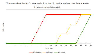
A microwell or microtitre plate typically consists of 6, 24, 96, 384 or more sample wells arranged in a 2:3 rectangular format. Most common laboratory microwell, is designed for working with nearly 0.5 ml of reactions.
The antigen / antibody binding to plate is mainly dependent on the temperature and duration. The most common method for coating plates involves adding a 5 +/- 3 μg/ml solution of protein dissolved in an alkaline buffer such as phosphate-buffered saline (pH 7.4) or carbonate-bicarbonate buffer (pH 9.4) at 4C for overnight. The binding can be increased by use of agents such as Gluteraldehyde or carbodiamide. For binding of high carbohydrate containing molecules Poly-L-lysine has been used. Modern industrial standards use more complicated chemistry to achieve higher quality. Plastic solid phases have an affinity to bind variety of proteins and can lead to increase in non specific reactions. This leads to background noise. This can be reduced (not eliminated), by blocking non specific sites on the plate.
Many different stabilizers are available at the manufacturer's disposal. Most of them are patented synthetic formulations that preserves the plate form drying.
The second most important reagent for ELISA is a conjugate. This is a marker antibody, that has got Enzyme on its Fc portion. Recall from the structure of antibody that the Fab portion is involved in binding the antigen and the Fc is free. The primary question here is how do you link the Enzyme to Fc tail of an antibody. The trick is to use linkers. Two broad range of linkers are available. Homolinker and Heterolinker. The type of linker to be used depends on the enzyme and antibody to be linked. Adipic Acid Dihydrazide, Glutaraldehyde, Periodate, N-succinimidyl 3-(2-pyridyldithio) propionate (SPDP) etc are used as linker for HRP. HRP (Horse Raddish Peroxidase) is a commonly used enzyme. Other choice of enzymes used include Alkaline Phosphatase, Acid Phosphatase, etc.
Enzyme
|
Substrate
|
Final absorbance and color
|
Alkaline Phosphatase
|
PNPP (p-Nitrophenyl Phosphate, Disodium Salt)
|
405 nm; Yellow
|
Horse Raddish Peroxidase
|
ABTS (2,2'-Azinobis [3-ethylbenzothiazoline-6-sulfonic acid]-diammonium salt)
|
410 nm; Green
|
TMB (3,3′,5,5′-tetramethylbenzidine)
|
450 nm; Yellow
|
Table 2: Enzymes and substrates used in ELISA
Reactions using peroxidase enzymes (such as HRP), on TMB substrate produces a soluble blue product. The color fades in short time. If intended to keep for longer duration with color preserved (especially in techniques such as Cell ELISA) ammonium heptamolybdate, Osmium tetroxide or nitroferricyanide can be added to stabilize the TMB chromophore.
In indirect ELISA, antibody to be detected is added to well containing coated antigen. If antibody of interest binds specifically, the antibody immobilizes via antigen to the solid surface. A second antibody conjugate that recognizes the Fc portion is added. If there is a stable immobilized Ag-Ab-Ab complex, the enzyme will be available for reaction after the washing step. The substrate will be converted to colored product. Finally the reaction is stopped using a stop solution. The final color is read by using a ELISA reader. Indirect ELISA is the method of choice to detect the presence of serum antibodies, such as HIV antibody.
 |
| Fig 4: Types of ELISA. Source |
In the sandwich technique, Antibody is immobilized to the well. The antigen in test, is allowed to bind. A second antibody (Conjugate), that binds to the Antigen-Antibody complex is used as marker/tracer. The stable, immobilized Ab-Ag-Ab will contain the enzyme to convert substrate as in Indirect technique. Sandwich technique is used to demonstrate antigens such as HBsAg in serum.
Theoretically, the technique of ELISA gives a higher sensitivity, as the enzyme can convert more substrates to colored product, provided the reaction can be allowed to proceed for a longer time. However, too much of incubation may enhance noise to signal ratio.
- Reverberi R , Reverberi L. Factors affecting the antigen-antibody reaction. Blood Transfusion. 2007; 5: 227-240. Link
- David Goldblatt. Affinity of Antigen–Antibody Interactions. els. 2001. Link
- A. Ansari, N.S. Hattikudur, S.R. Joshi, M.A. MedeiraELISA solid phase: Stability and binding characteristics. Journal of Immunological Methods. Volume 84, Issues 1–2, 28 November 1985, Pages 117–124. Link
- A novel surface treatment to reduce non-specific binding to microplates. Link
- Overview of ELISA. Link


Comments
Post a Comment